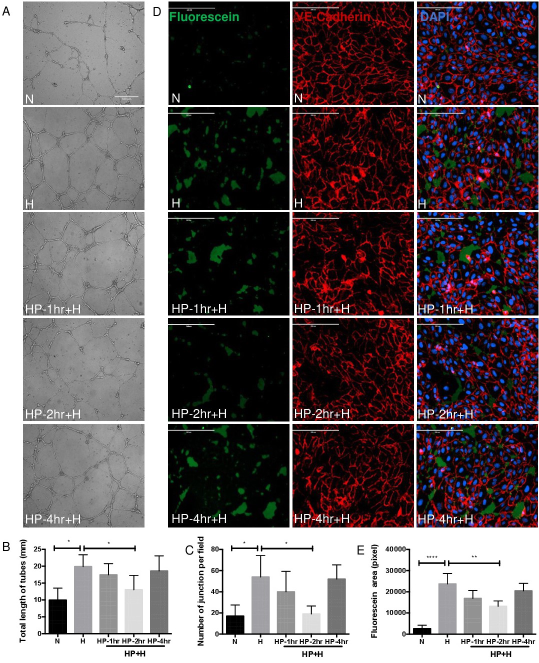
Fig. 5. Hypoxic preconditioning decreases HIF-1-dependent up-regulation of angiogenesis and vascular permeability. (A-C) Stimulation of HRMECs tube formation by conditioned media from MIO-M1 cells exposed to 1% O2 hypoxia 48 hours (H) compare to 20% O2 normoxic condition (N). Supernatant from HP-2 hours (HP-2hr) reduced both the total length and junction number of tubes compared to Hypoxic group (n=12 per group). Images was performed with Leica DMI6000 microscope (x5). Scale bar, 500 μm. (D, E) HUVECs were plated on coverslips coated with biotinylated gelatin and grown to confluence. then treated for 10 min with conditioned medium from cultured muller cells. FITC-avidin was added for 3 min, cells were fixed and subjected to immunofluorescence staining for VE-cadherin (red) to visualize cell-cell contacts. Green fluorescence depicts areas permeable for FITC-labeled avidin. Conditioned medium from hypoxic MIO-M1 cells increase area of FITC-positive intercellular spots and decreased by HP-2 hours (n=3 per group). Scale bar, 200 μm. Data represents means ± SD of relative values vs control from 3 independent experiments. *P<0.05; **P<0.01; ***P<0.001; ****P<0.0001, statistical analysis was performed with one-way ANOVA with Dunn's test for multiple comparisons.